We have invested in advanced technology called

OCT that allows our eye doctors to have a clearer image of your eye.

Maestro2

Ocular Coherence Tomography

Optical coherence tomography (OCT) scans are important for establishing early diagnoses for many serious eye diseases. Learn why we have invested in advanced technology for the care of your eyes and vision.

When should I have an OCT scan?

If you are over 25 years of age or at risk of developing ocular diseases, your comprehensive eye examination may include an OCT scan. OCT scans enable your doctor to compare the images from previous years in order to detect any abnormalities or thickening of the retinal layers.
The information provided by an OCT scan will also be useful for monitoring your treatment if you have been diagnosed with ocular disease. In case you have any questions about the OCT exam, do not hesitate to reach out to your nearest Amplify EyeCare practice either via a call or in-person visit.
The optometrist may recommend an OCT scan if you have any of the following eye diseases or family history of eye disease.  There are many conditions that may benefit from an OCT scan. Below we provide a more comprehensive list.

Diabetes or diabetic retinopathy

Glaucoma

Retinal Detachment

Age Related Macular Degeneration

Why should you schedule an eye exam with an optometrist that has an OCT?

An OCT is one of the most expensive technologies in a modern optometry office, ranging in price from $30,000-$80,000. When an optometry office has invested in this equipment it is a strong indication of the office's commitment to the healthcare of their patients. Furthermore the benefits of an OCT in allowing for a baseline of your eyes is important as you age, with every visit your eye doctor is able to look at previous scans and assess for any change. For patients with eye disease the benefits of an OCT are exceptionally high, the high resolution 3D images allow the optometrist to identify early signs of disease progression before permanent vision loss occurs.
The Ocular Coherence Tomography (OCT) technology is available in these Amplify EyeCare locations:

What Is OCT?

Optical coherence tomography (OCT) provides detailed images of the layers in the optic nerve and retina. In the same way as an ultrasound, OCT uses light waves rather than sound waves to direct focused light beams into the eye. The beams then get reflected out from the eye. After processing, eye care professionals can view detailed cross-sectional images of various structures within the eye. An OCT takes roughly 15 minutes to complete. It is an easy and non-invasive test.

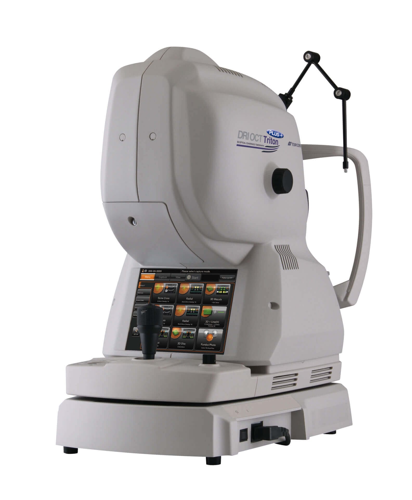
The OCT test gives highly detailed and accurate 3D imaging of the inner workings of your eye while being super comfortable for the patient. The OCT is: 

  • noninvasive 
  • painless 
  • minimal health risks
  • zero radiation

What is the use of optical coherence tomography?

Retinal conditions

Optic nerve disorders

Pre and post-surgical assessment

Biomedical Imaging Using Optical Coherence Tomography

Retinal conditions

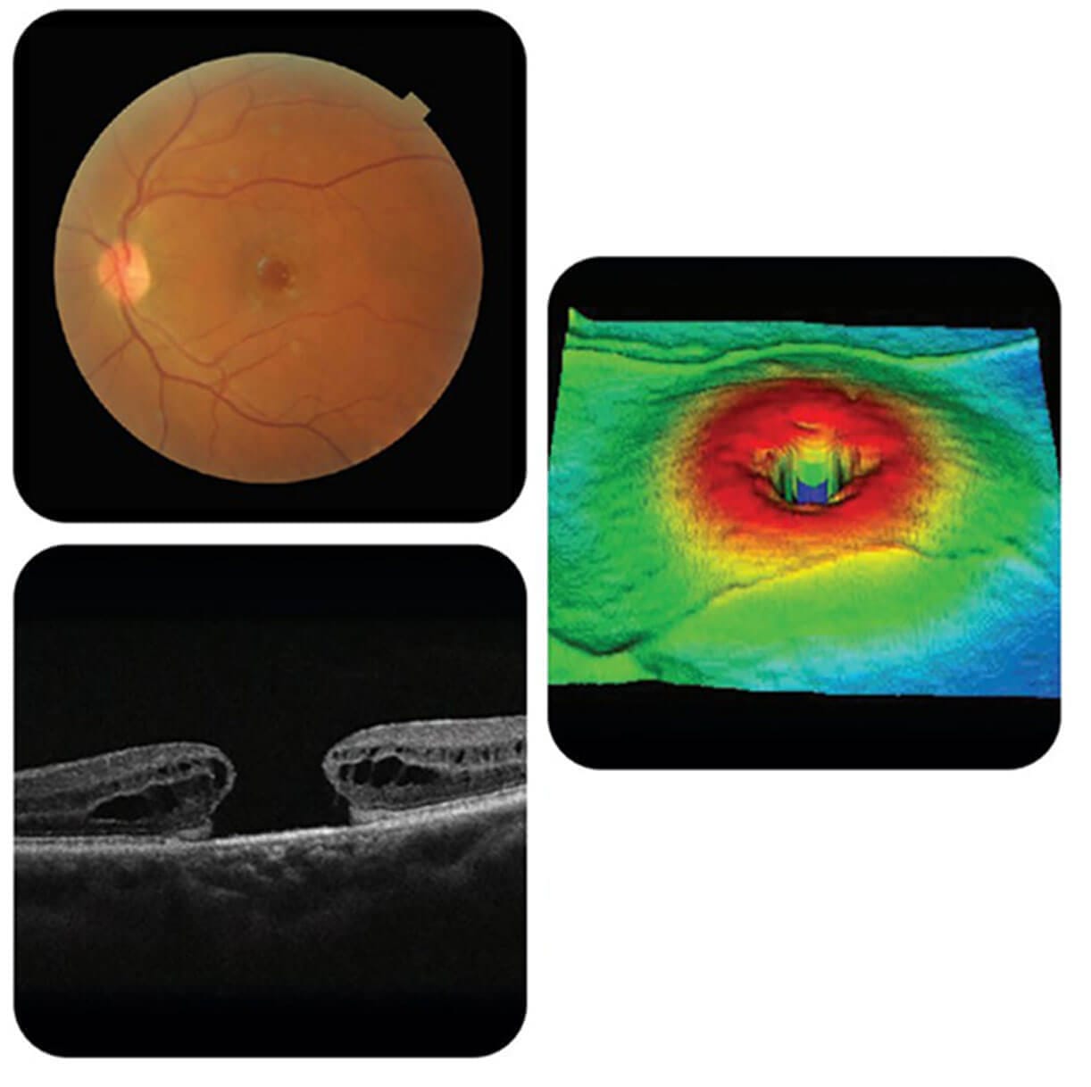
Many retinal conditions can be diagnosed with OCT. It is generally easier to image lesions in the macula than in the mid and far periphery. Among the conditions OCT is most useful for diagnosing are:

  • Macular hole
  • Macular pucker/epiretinal membrane
  • Vitreomacular traction
  • Macular edema and exudates
  • Detachments of the neurosensory retina
  • Detachments of the retinal pigment epithelium (e.g. central serous retinopathy or age-related macular degeneration)
  • Retinoschisis
  • Pachychoroid
  • Choroidal tumors
  • Subretinal fluid

It is possible to diagnose certain conditions using only an OCT (e.g. macular hole). For other disorders, particularly vascular disorders of the retina, additional tests may be necessary (such as fluorescein angiography or indocyanine green angiography).

Optic nerve disorders

OCT is becoming increasingly popular for evaluating retinal nerve fiber layer and ganglion cell layer thickness to diagnose optic nerve disorders:

  • Glaucoma
  • Optic neuritis
  • Non-glaucomatous optic neuropathies
  • Alzheimer's disease
Pre and post-surgical assessment

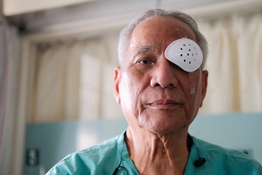
Cataract surgery is one of the most common surgeries performed in the US, the OCT is an incredibly powerful tool in pre operative and post operative care for cataract patients. 

Utilizing OCT imaging as a tool for pre and postoperative assessment can provide invaluable information for the surgical management of macular holes and retinal detachments. By using OCT, surgical outcomes can be visualized, confirming reattachment and normal contour. Post-surgical imaging can sometimes be challenging because of reduced OCT signal strength caused by ocular turbidity; however, images are adequate for subjective, if not quantitative, interpretation. In cases of poor ocular media, pre-surgical scanning can reveal pathologies that can complicate surgery, such as an undetected macular hole, choroidal neovascularization, edema, or vitreomacular traction syndrome. Due to the OCT's scanning beam technology, it is possible to image even through a small pupil or tiny peripheral opening in a dense cataract that otherwise would obstruct a thorough examination.

Biomedical Imaging Using Optical Coherence Tomography

OCT has several characteristics that point to its future importance as biomedical imaging technology.

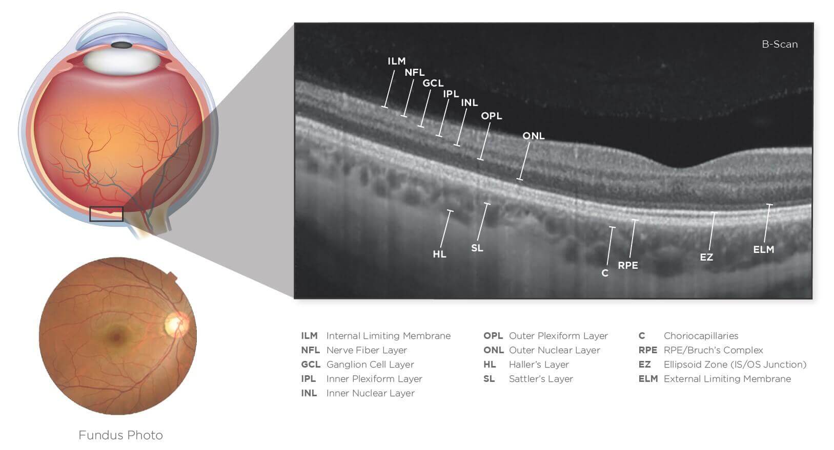
OCT images can have axial resolutions of one to fifteen micrometers, which is a significant increase over conventional ultrasound. Contrary to ultrasound, imaging can be performed directly through air, without direct contact with the tissue or a transducing medium.

Imaging can be performed in situ, without the need to excise a specimen. By doing so, it is possible to image structures where a biopsy would be dangerous or impossible. As a result, it also allows better coverage, reducing the sampling errors associated with excisional biopsy.

In contrast to conventional biopsy and histopathology, imaging can be performed in real time without the need to process a specimen. In this way, pathology can be monitored on screen and stored as high-resolution videos. By coupling real-time imaging with surgery, it is possible to provide surgical guidance based on real-time diagnosis.

Time-domain versus spectral-domain OCT in Age Related Macular Degeneration

The new spectral-domain (SD) OCT devices use a spectrometer in the receiver to analyze the spectrum of reflected light on the retina and calculate the depth of the structures, according to the fourier principle. The time-domain (TD) OCT uses this technology to eliminate the need for mechanically moving the reference arm, thereby increasing the speed of image reception and axial resolution. SD-OCT uses different algorithms than TD-OCT, and the retinal thickness measurements are not comparable between the two. SD-OCT also offers a faster rate of capturing images and higher definition. In contrast to TD-OCT which measures retinal thickness from the internal limiting membrane to the highest hyperreflective band, i.e., that combines the inner and outer segments of the photoreceptors, SD-OCT measures this near the RPE. As a result, SD-OCT measures retinal thickness values much higher than that obtained by TD-OCT.

Contact Us To Amplify Your EyeCare

phone-handsetcalendar-fullarrow-up linkedin facebook pinterest youtube rss twitter instagram facebook-blank rss-blank linkedin-blank pinterest youtube twitter instagram